
Nevi (Moles) – Hazard Criteria
Any alteration of the skin should be checked by a specialist dermatologist for if there are certain features that internationally referred to as ABCD (the first four letters of the Latin alphabet).
- Asymmetry – Asymmetry (The Left Side Is Different From The Right)
- Border Irregularity – Irregular Boundaries (The Boundaries Of The Nevus That Is Not Clear In Relation To The Surrounding Skin
- Colour Variation – Color Variety (The Nevus Has Not Been Uniform Coloring, But Contains Various Colors As Light And Dark Brown, Black, Pink, White And Blue)
- Diameter Over 6 Mm – Diameter (Melanoma Is Usually Larger Than 6 Mm In Diameter)
- Change In The Size, Shape Or Color Of A Nevus
- The Nevus Bleeding Or Secrete Liquid
- The Nevus Causes Itching, It Is Hard To The Touch, Without Form, Is Swollen Or In Pain
- A New Nevus Occurs In Clean Skin\
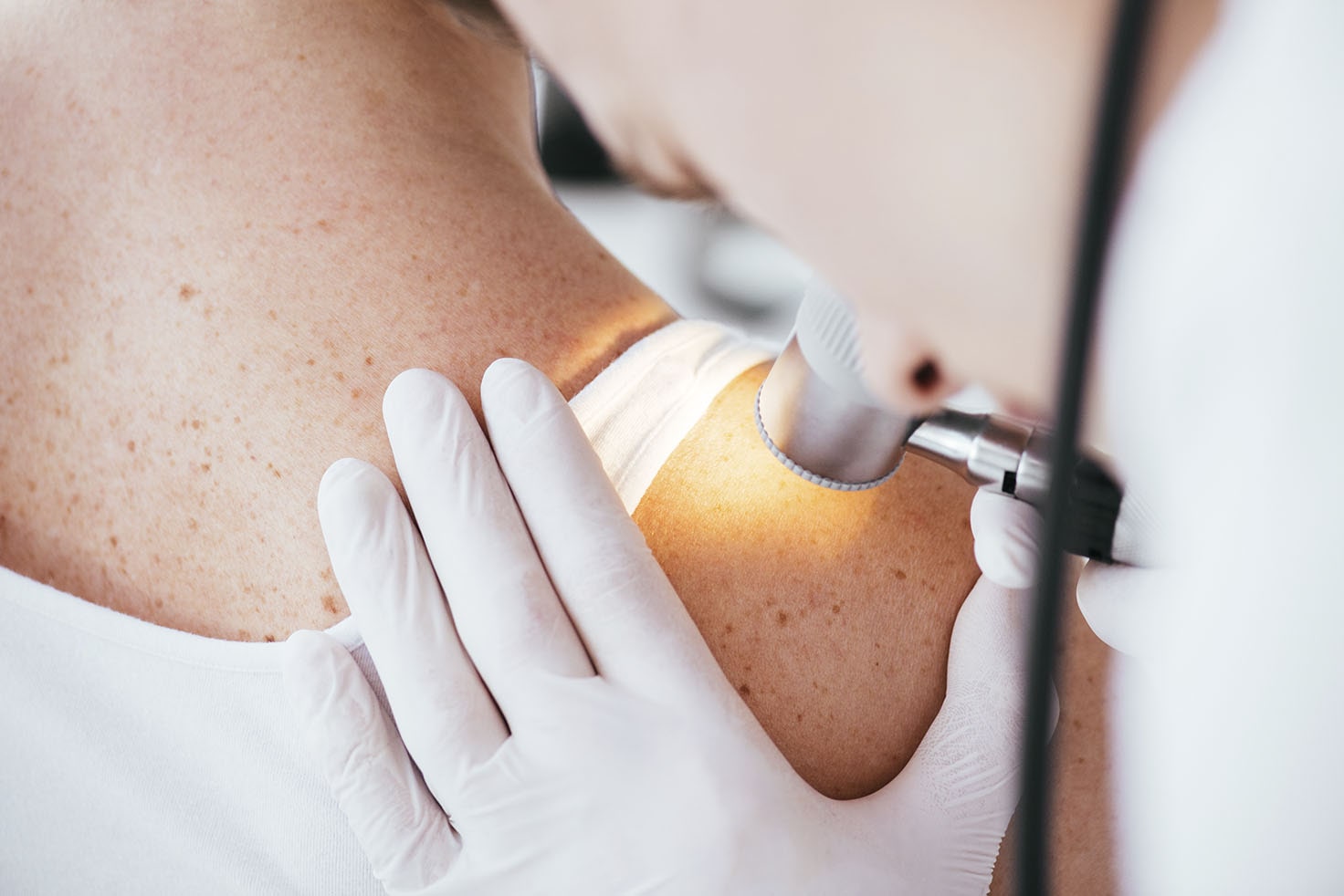
What Is The Digital Mole Mapping;
It is an extremely important consideration for any moles that there are documented photographs of the olives (stored in the machine but are given and the patient) and to be able to identify any future changes.
Digital Δερματοσκόπιο is the most specialized and reliable device for the diagnosis of Skin Cancer. The special camera δερματοσκοπίου magnify the olive tree in order to observe items that can not be seen by the human eye. The images from each mole of the body are recorded, mapped and stored in an individual file. With a special computer program finally, an analysis of each nevus and exported to the amount of risk that helps in the final decision for removal, or monitoring of the nevus. Even for aesthetic reasons if you are going to be a removal of an olive tree, the examination is necessary prior.
The examination is painless, harmless, without radiation and is suitable even for children.
Digital Δερματοσκόπιο MicroDERM
The first Digital Δερματοσκόπιο 2nd generation in accordance with the FDA, the microDERM, it helps the physician and the patient using the visual of a computer. The first principle of the neural networks – αυτοδιδασκόμενες machines that were developed with the human mind as a model. This technology will redefine the handling of information and the diagnosis of the findings. The keys for the professional, systematic, timely diagnosis of cancer of the skin is the automation and standardization, in order to give greater quality and better efficacy in the treatment of each patient. The new techniques of light and optics allow for better images. The επιμικροσκοπικές downloads are complemented by a three-dimensional image of the skin lesion and create an impressive, high-resolution and quality image. This allows a proper assessment of skin lesions. The patented optical precision rejects the creation of colour and light variations, and morphological deformities. Such mistakes occur on machines of the first generation of βιντεοδερματοσκοπίας that were simplistic methods of growth, and prevent the accurate assessment of skin lesions. The camera system of the microDERM covers this need and more. The built-in microprocessors that offers the microDERM, automatically adjust the intensity of the light and the composition of the color so that it remains the same, which allows for excellent and stable quality at every stage of growth.
The microDERM is not just a registration system, but rather represents the new generation of medical equipment in a timely and rapid diagnosis of skin cancer. The ever-increasing incidence of malignancies and mortality of cancer of the skin, make the system necessary.
MicroDERM Hair Analysis Hair
For the diagnosis of disorders of the hair ( p.x. loss of hair ) are necessary for the physiological parameters of the hairs. The oldest method for the τριχογράμματα was cutting about 100 hairs and their examination under a microscope. Today there is the φωτοτριχόγραμμα, a non-invasive method for the estimation of the parameters of the hair, the method of which is contained in the microDERM. The microDERM goes even further as it has to the doctor who is handling the digital βιντεοτριχόγραμμα, a specialized system of analysis of hair through a computer, which is designed for the analysis of changes in the growth of each hair. In this way, the microDERM can often replace the classic τριχόγραμμα and be used for all types of disorders of the hair.